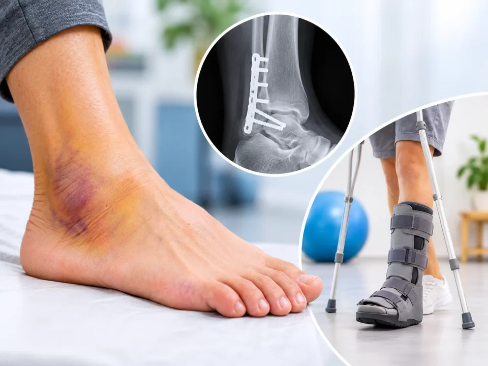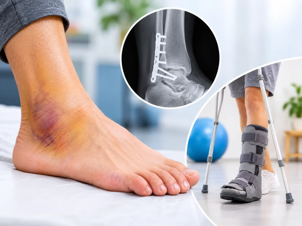

When Does an Ankle Ligament Tear Require Surgery?
October 20, 2026
Shoulder Pain When Raising Arm Causes Relief and Recovery
November 3, 2026

An ankle that keeps hurting long after a sprain may have an ankle cartilage injury. A common form is an osteochondral lesion of the talus: damage to the joint surface and the bone beneath it. Cartilage heals slowly, so the ache lingers. An MRI confirms the diagnosis. Treatment ranges from protection and physiotherapy to keyhole surgery.
You rolled your ankle weeks ago. The swelling went down and the bruise faded. Yet the ankle still aches on stairs, or clicks when you pivot on the court. The outside has healed. Something inside the joint may still be hurting, and that something is often cartilage.
Why Does My Ankle Still Hurt After a Sprain?
A simple sprain stretches ligaments, and these usually settle within a few weeks. If pain, swelling or clicking continues beyond that, the same twist may also have bruised or cracked the joint surface. Ligament pain can mask this deeper injury early on. As the ligament settles, the deeper ache becomes clear. Understanding the possible complications of an ankle sprain can help explain why some ankle injuries cause symptoms beyond the usual recovery period.
Other causes of lingering pain exist too, including loose ligaments and tendon injury. That is why a proper assessment matters.
Why Cartilage Heals So Slowly
Cartilage is the smooth, glossy layer that covers bone ends inside a joint. In the ankle, the talus (the bone that sits between the shin and the heel) carries a large share of your body weight. Healing needs blood. Cartilage has no blood supply of its own, and the talus has a limited supply too. A damaged patch struggles to repair itself.
Cartilage also has no nerve endings. The ache you feel comes from the bone underneath and the joint lining, which react when the surface is damaged.
Ankle Cartilage Damage Symptoms
Patients rarely say “cartilage”. They use everyday words:
- A deep ache inside the joint that rises with walking, running or jumping
- Pain that returns after a long day on your feet
- Swelling that comes and goes with activity
- Clicking, catching or a brief locking feeling
- Stiffness after sitting, or a sense the ankle may give way
- Pain that continues after a normal X-ray
Ankle clicking and popping can have different causes. Read more about ankle cracking and popping and what these sounds may indicate.
The Where-When-What check
Three questions help you describe your pain clearly.
- Where? Deep in the joint, often at the front, inner or outer side of the ankle.
- When? It builds with load and eases with rest.
- What? The ankle clicks, catches, swells or feels weak.
Two or three matches justify an assessment. The check helps you describe symptoms. A specialist gives the diagnosis.
Sprain, Fracture or Cartilage Injury: How They Differ?
These injuries can happen together, but each has its own pattern.
| Feature | Ligament sprain or instability | Ankle fracture | Cartilage and bone injury of the talus |
| Typical start | Twist or roll | Fall, heavy twist or impact | Sprain, fracture or repeated stress |
| Pain pattern | Tender around the ligament; eases over weeks | Sharp; often hard to bear weight | Deep ache; linked to load; persists |
| Common extras | Looseness, repeated rolling | Marked swelling or deformity in some cases | Clicking, catching, swelling after activity |
| Key tests | Examination; MRI at times | X-ray; CT at times | MRI; CT at times |
Our pages on ankle ligament reconstruction and ankle fracture treatment cover the first two injuries in depth.
What Is an Osteochondral Lesion of the Talus?
An osteochondral lesion of the talus is damage to cartilage and the bone directly beneath it, on top of the ankle bone. “Osteo” means bone. “Chondral” means cartilage. The injury may be a bruise, a crack, a loose flap or a small fragment that has come away. Some lesions form fluid-filled pockets in the bone, called cysts.
Surgeons often grade lesions using a system first described by Berndt and Harty. It looks at whether the fragment is compressed, partly detached or fully loose. Grade, size and location all guide treatment.
How an Ankle Cartilage Injury Is Diagnosed?
Diagnosis starts with your story and a hands-on examination. Imaging follows. An ankle X-ray can look normal while the joint surface is damaged. MRI shows the cartilage and any swelling in the bone below. A CT scan shows bone detail, including cysts and loose fragments. For patients with persistent symptoms, specialist assessment can help determine the appropriate approach to cartilage injury treatment.
At OrthofootMD, imaging can include digital radiography and 3D imaging. Intraoperative fluoroscopy guides surgery when it is needed.
Ankle Cartilage Injury Treatment: From Gentle to Surgical
Treatment depends on lesion size, depth and location, plus your age and activity goals.
Protect and rebuild
Many people begin here. This means activity changes, a supportive boot or brace for a period, and physiotherapy to restore balance, strength and movement. It can suit smaller, stable lesions, and a trial period shows how the ankle responds.
Biologic injections
Platelet-rich plasma (PRP) is a concentrate made from your own blood. Other regenerative injections also exist. Evidence for ankle cartilage is still developing, so they may ease symptoms while other treatment continues. Your specialist explains the current evidence for your situation.
Keyhole surgery
When symptoms persist, ankle arthroscopy lets the surgeon see inside the joint through small cuts. A tiny camera guides the work. Loose fragments can be removed and damaged tissue cleaned.
Bone marrow stimulation is a common addition. Tiny holes in the bone under the lesion encourage the body to form repair tissue called fibrocartilage. This tissue differs slightly from original cartilage, which is why lesion size matters when choosing this option.
Restoring the joint surface
Larger lesions, cysts or earlier failed surgery may call for more. Options include fixing a fragment back in place, bone grafting, or transferring cartilage and bone from another area or a donor. Cell-based and scaffold techniques also exist. Suitability varies, so the right choice emerges after imaging and discussion.
The Road Back to Walking, Running and Sport
Recovery follows stages: protect the ankle, restore movement, rebuild strength, then return to impact. Timeframes vary with the treatment and the lesion. Your surgeon gives you a plan for yours.
Runners often return through a graded walk-run plan. Soccer players add cutting, pivoting and landing drills before match play. A sports-specific recovery pathway matches those steps to your sport. After surgery, weight-bearing rules differ between patients. Follow yours closely, because early overload can affect how the repair settles.
While You Wait for an Assessment
Ease off high-impact activity such as running, jumping and pivoting. Choose supportive footwear. Stop an activity if it brings on a deep ache or a catching feeling. Seek urgent care for severe swelling, visible deformity or an ankle that cannot bear weight. Those signs point to a different problem, such as a fracture.
When to Book an Assessment
Consider seeing a foot and ankle specialist if you notice:
- Pain or swelling well beyond the usual sprain recovery time
- Clicking, catching or locking
- Repeated giving way
- Ongoing pain after a normal X-ray
- Ankle pain that limits running, work or stairs
Dr Gowreeson Thevendran is a triple-fellowship-trained orthopaedic and trauma surgeon. He consults at Mount Alvernia Hospital and Mount Elizabeth Novena Specialist Centre in Singapore. His practice focuses on foot and ankle conditions, with an emphasis on keyhole surgery and biologic therapies.
If your ankle cartilage injury is holding you back, schedule an assessment with our foot and ankle specialist.
FAQs
Look for a deep ankle ache that builds with activity, along with clicking, catching or swelling that lingers after a sprain. Only an examination and imaging, usually MRI, can confirm it. A foot and ankle specialist can arrange both.
Mild sprains often settle within a few weeks. Severe sprains can take longer. Pain that persists well beyond the expected window, or that changes to a deep, load-related ache, deserves a specialist review.
Small bruises of cartilage and bone may settle with protection and rehabilitation. Cartilage itself repairs poorly because it lacks a blood supply. Some lesions improve; others persist and need further treatment.
Larger lesions can appear on X-ray, but early or small ones are often missed. MRI shows cartilage and bone swelling clearly. CT adds bone detail when surgery is being considered.
They are different conditions. A lesion is damage to a localised area of the joint surface. Arthritis involves wider wear. Persistent damage may raise the risk of wear over time, which is one reason to assess it early.
Continued impact on a damaged joint surface can aggravate symptoms. Lingering pain deserves assessment before you resume full training. Your specialist can advise on what load the ankle can currently handle.
Many patients begin with protection, physiotherapy and sometimes injections. Surgery becomes an option when symptoms persist, or when imaging shows a loose fragment or a larger lesion.
Recovery depends on the procedure, the lesion and your sport. It follows stages from protection to full impact. Your surgeon sets a timeline after reviewing your scans and surgery details.
PRP is used to ease symptoms in some patients. Research in ankle cartilage is still developing, so results vary. Your specialist can explain whether it suits your lesion and goals.
A twisting injury is a common cause, including sprains and fractures. Repeated stress from running or jumping can also damage the joint surface over time. Sometimes the cause stays unclear. The result is injury to the cartilage and the bone beneath it, which imaging confirms.
A foot and ankle orthopaedic surgeon is the usual choice for pain that lingers after a sprain. They examine the joint, review MRI or CT scans and explain the non-surgical and surgical options.